The following resources are designed for use by clinicians and /or health service providers to assist with the implementation of services to manage the increasing number of patients presenting with difficult intravenous access (DIVA).
DIVA Resources for Clinicians & Organisations
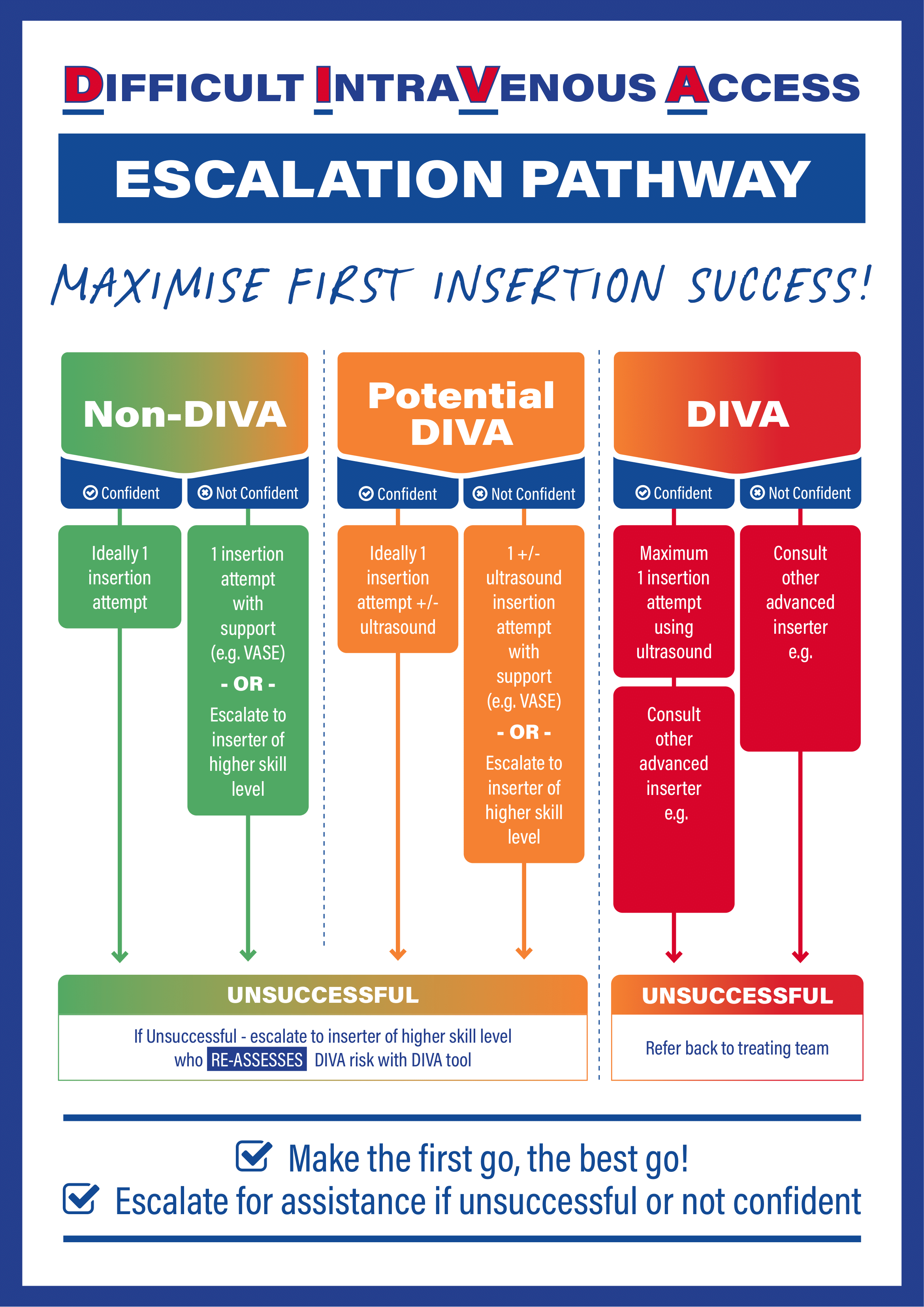
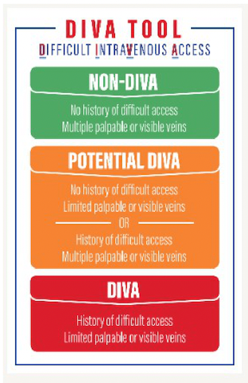
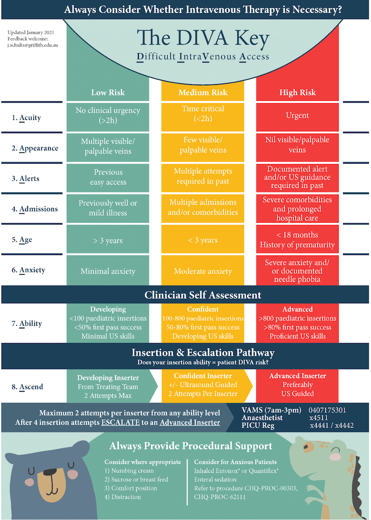
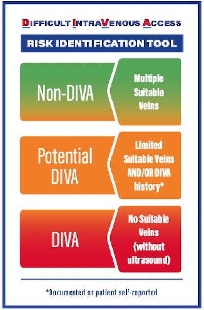
Identification of DIVA
The recent Australian NHMRC funded DART 3 study co-developed DIVA identification and escalation pathways with the three participating hospitals, these can be found here:

More information about the DART 3 study can be found here and the published protocol is available here.
Ultrasound use in PIVC Insertion in DIVA Patients
Ultrasound can be an effective assistive technique in peripheral intravenous catheter insertion, especially in patients with difficult intravenous access (DIVA). Ultrasound can differentiate between structures such as bone, nerve bundles, muscle and veins and arteries, showing the operator images in real time, including the ability to see the needle when inserting the peripheral intravenous catheter. There are many types of ultrasounds ranging from expensive large machines to less expensive small handheld probes that connect to tablets the size of a mobile phone. Some companies who specialise in ultrasounds are: https://www.philips.com.au/healthcare, https://www.sonosite.com/au and https://www.gehealthcare.com.
Ultrasound guided peripheral intravenous catheter (USPIVC) insertion is a specialist skill that requires education and training and most of all, opportunities for the inserter to maintain their competence and confidence in the skill. There are several training resources available for USGPIVC insertion, generally these include a theory component followed by a face-to-face, hands-on competency development workshop.
Following training, competence of the skill is often required via skills assessment, examples of skill assessments are available below.
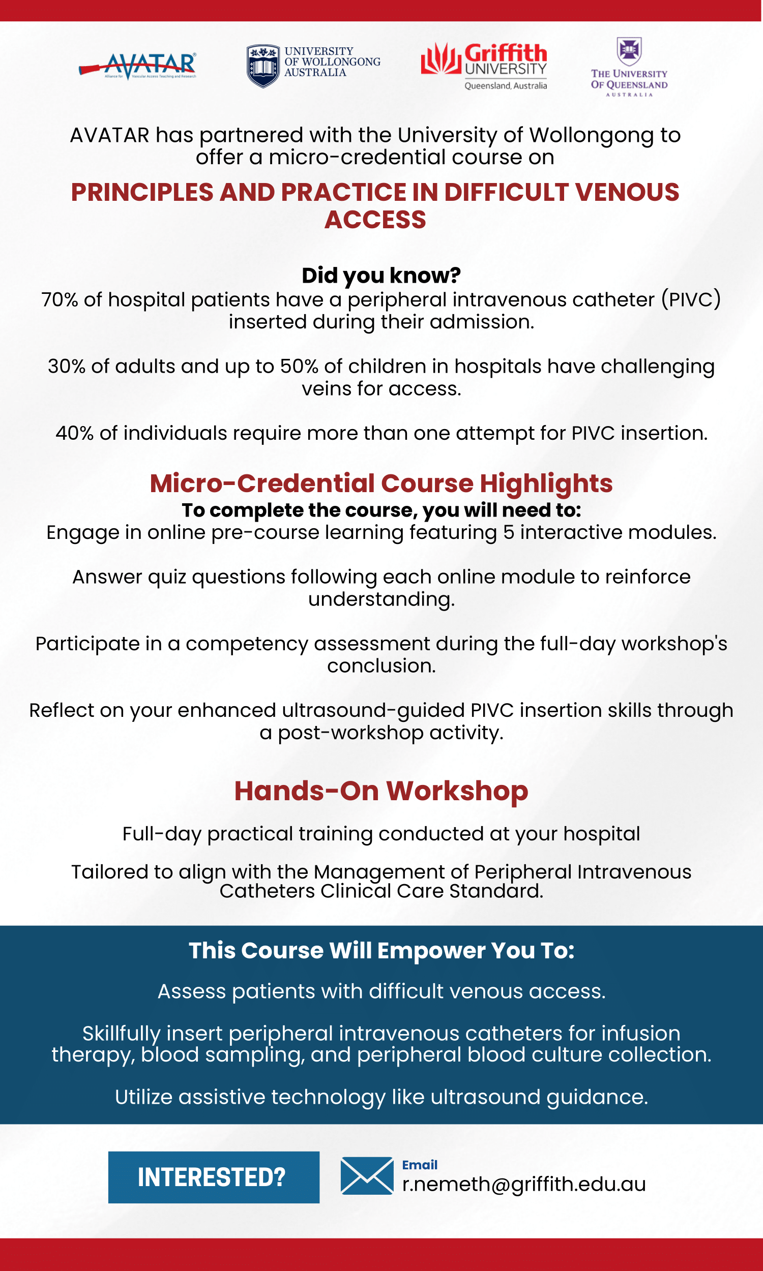
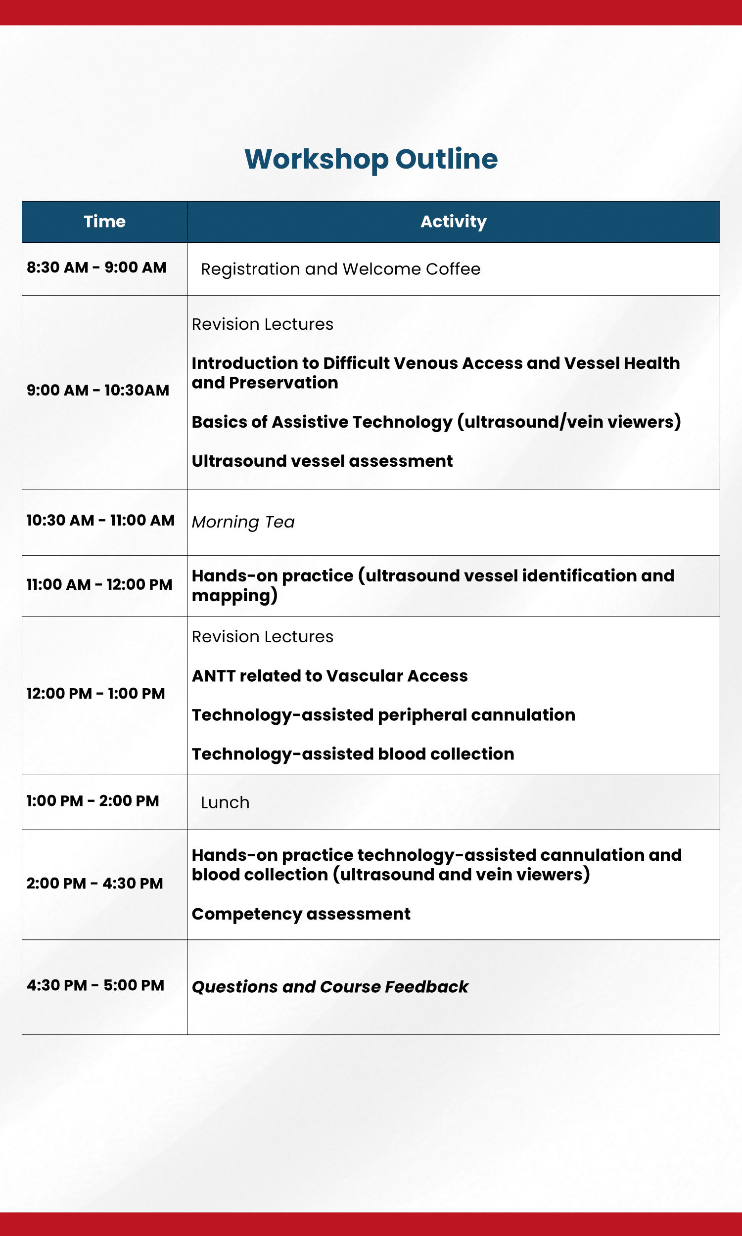
The University of Wollongong (UoW) offers a micro credentialed course, Principles and Practice in Difficult Venous Access, information is available here. AVATAR has partnered with UoW to offer this course at local health services anywhere in QLD, more information is available below.
Queensland Health’s Clinical Skills Development Service also offers an USGPIVC course aimed at healthcare workers who are already competent in PIVC insertion and would like to develop USGPIVC skills. Course information can be found here, this course includes free online theory.
Following training, competence of the skill is often required via skills assessment, examples of skill assessments are available below.
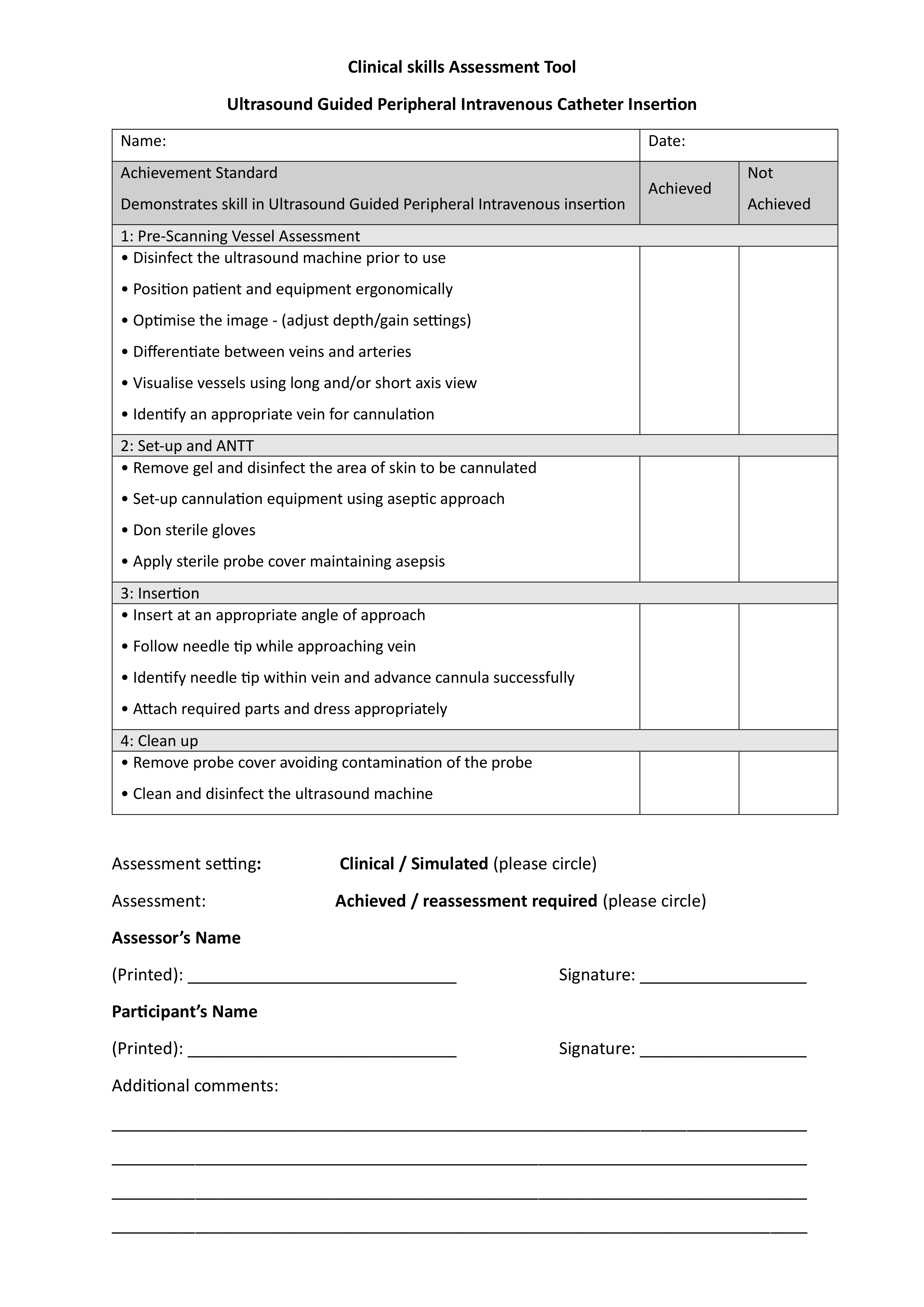
Brief Skill Assessment USGPIVC Example
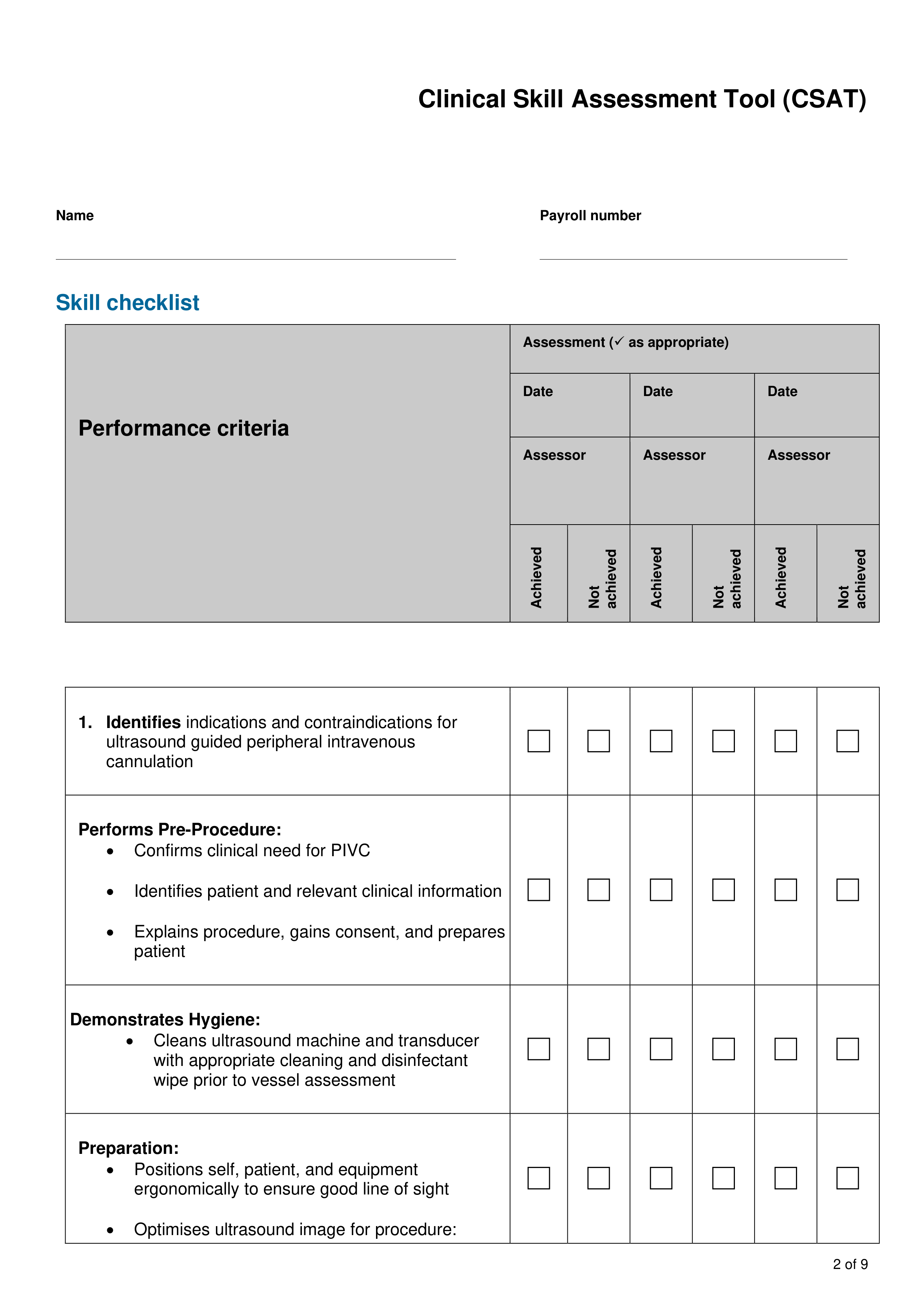
Extended Skills Assessment USGPIVC Example
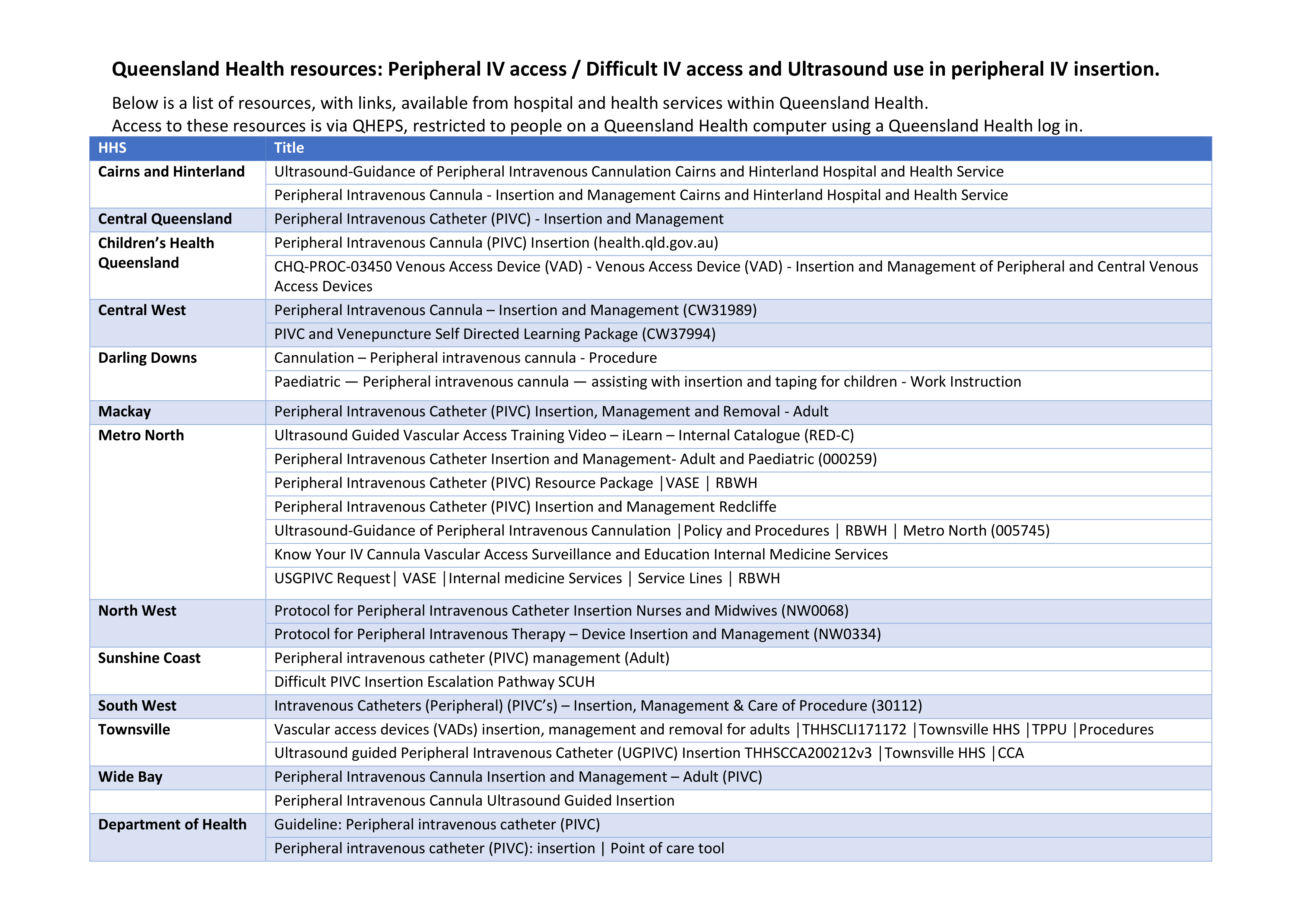
Queensland Health Hospital and Health Services have many policies and procedures on Peripheral IV access, DIVA and ultrasound use in PIVC insertion, a list of these is below, these are only accessible with Queensland Health access from a Queensland Health computer.
Using aseptic non-touch technique when inserting the peripheral intravenous catheter is essential. This includes disinfection of the Ultrasound probe before and after each use. There are several products and systems used by organisations, some recent literature has evaluated the difference between disinfection wipes, here and the journal of ultrasound in medicine has published a guidance paper on cleaning ultrasound transducers, here.
Some short videos’ can be found below, these show the use of ultrasound in vein assessment and PIVC insertion.
Basilic Vein
Vein Assessment
Longitudinal Approach
Other Assistive Technologies used in PIVC insertion in DIVA patients - Near Infrared:
Near infrared (NIR) is another type of technology to visualise veins. Presently, VeinViewer® is the only product in this category in Australia. NIR may be more appropriate than ultrasound for occasional users, in children or if only superficial veins are being targeted. The metrics that can be
assessed using NIR include:
- Real size, shape, and location of the veins
- Gauge of depth
- Blood flow
- IV flushing; assessment of patency/infiltration
- Bifurcations and Valves
- Catheter Sizing
The depth of penetration of VeinViewer® is up to 10 mm deep for peripheral veins and up to 15 mm deep for blood patterns (ie: hematomas, previous IV sites). Depth of visualisation is dependent on the anatomy and condition of each patient.
Watch a short video here or for more information visit https://christiemed.com/
Literature
Recent Research Papers on the Background of DIVA
Defining difficult intravenous access (DIVA). A systematic review
Recently Published Research about the Management of DIVA
Handheld Ultrasound Devices for Peripheral Intravenous Cannulation
A clinical pathway for the management of difficult venous access
Recently Published Paediatric Specific DIVA Studies